Tendon Diagram - Peroneal Tendon Syndromes Practice Essentials Epidemiology Functional Anatomy - Foot anatomy diagram, foot joint diagram, foot sprain diagram, foot tendons and ligaments pain, leg tendon diagram.. Jul 01, 2021 · free body diagram for calculating deltoid force. The achilles tendon connects the heel to the calf muscle and is essential for running jumping and standing on the toes. The anterior cruciate ligament prevents the femur from sliding backward on the tibia (or the tibia sliding forward on the femur). For images of the muscle, click on each link under location. Foot anatomy diagram, foot joint diagram, foot sprain diagram, foot tendons and ligaments pain, leg tendon diagram.
For images of the muscle, click on each link under location. Human hand tendon diagram (page 1) hand tendons diagram muscle blank drawing these pictures of this page are about:human hand tendon diagram the golgi tendon organ. A muscle's origin is where a tendon attaches it to the *less* movable bone. On the other hand, the insertion is where a tendon attaches that muscle to the *more* movable bone. Learn about the causes, symptoms, and treatment of wrist tendonitis here.
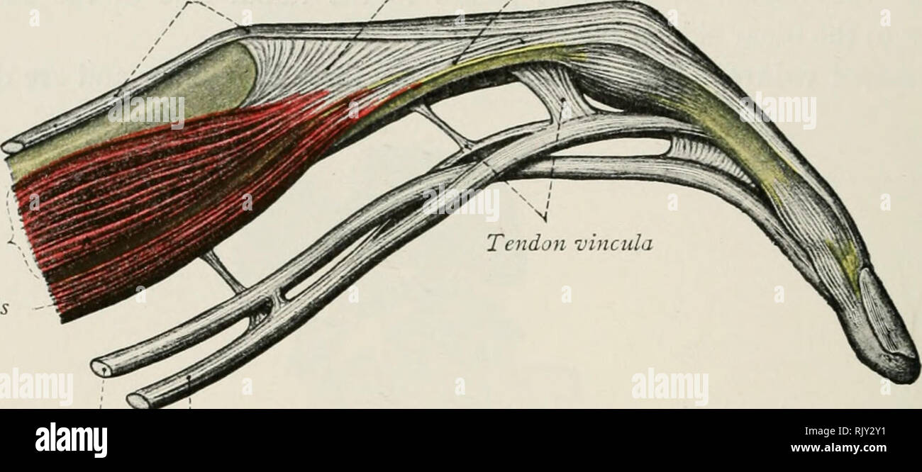
Atlas And Text Book Of Human Anatomy Anatomy Atlases 202 Atlas And Text Book Of Human Anatomy Tendon Of Extensor Digilorum Conun Tendon Of Interossnis Tendon Of Lumhricalis Interosscus Lumbricalis Tendon from c8.alamy.com Muscle of the frog by label The tendon is firmly connected to muscle fibres at one end and to components of the bone at its other end. The achilles tendon is also called the calcaneal tendon. The tendon runs down the back of your lower leg from the back of the knee to the heel. The achilles tendon attaches the muscles of the calves to the bones of the ankle and foot. A tendon, also known as a sinew, is a fibrous tissue that helps to facilitate this movement. In the back and elsewhere in the body, tendons attach muscles to bones. This tendon connects the patella (kneecap) to the tibia.
The achilles tendon connects the heel to the calf muscle and is essential for running jumping and standing on the toes.
Tendons attach muscles to bones. Brings leg back to and across body. If you are fortunate, you. If you tear the biceps tendon at the shoulder, you may lose some strength in your arm and have pain when you forcefully turn your arm from palm down to palm up. Ligaments join the knee bones and provide stability to the knee: A tendon, also known as a sinew, is a fibrous tissue that helps to facilitate this movement. For images of the muscle, click on each link under location. Tendons are found throughout the body, from the head and neck all the way down to the feet. The knee joint is a complex structure that involves bones. This tendon connects the patella (kneecap) to the tibia. On the other hand, the insertion is where a tendon attaches that muscle to the *more* movable bone. Diagram illustrating tendonitis and tendon rupture symptoms can vary from aches or pains and local joint stiffness , to a burning that surrounds the whole joint around the inflamed tendon. Foot and ankle musculoskeletal key :
A tendon, also known as a sinew, is a fibrous tissue that helps to facilitate this movement. For images of the muscle, click on each link under location. The anterior cruciate ligament prevents the femur from sliding backward on the tibia (or the tibia sliding forward on the femur). Ligaments join the knee bones and provide stability to the knee: The tendon runs down the back of your lower leg from the back of the knee to the heel.
What Are Muscles Tendons And Ligaments Examples Of Socratic from solidlifefitness.com Tendons are remarkably strong, having one of the highest tensile strengths found among soft tissues. The achilles tendon connects the heel to the calf muscle and is essential for running jumping and standing on the toes. Extends spine and trunk back. On the other hand, the insertion is where a tendon attaches that muscle to the *more* movable bone. Raises and rotates arm in all directions. Tendons are found throughout the body, from the head and neck all the way down to the feet. The achilles tendon is the strongest and largest tendon in the body. Tendons, located at each end of a muscle, attach muscle to bone.
The tendon is firmly connected to muscle fibres at one end and to components of the bone at its other end.
Human anatomy diagram from the back view 12 photos of the human anatomy diagram from the back view human anatomy diagram back view organs, human anatomy diagram rear view, human muscles, human anatomy diagram back view organs, human anatomy diagram rear view. Medical illustration of human arm muscles, veins and nerves. Human hand tendon diagram (page 1) hand tendons diagram muscle blank drawing these pictures of this page are about:human hand tendon diagram the golgi tendon organ. The achilles tendon is the strongest and largest tendon in the body. The cause is repeated contraction of the forearm muscles that you use to straighten and raise your hand and wrist. Possibly the most important tendon in terms of mobility is the achilles tendon. Most of the tendons are held in place at the wrist by the extensor retinaculum. Jul 05, 2018 · the foot diagram has a complex structure made up of bones, ligaments, muscles, and tendons. The achilles tendon connects the heel to the calf muscle and is essential for running jumping and standing on the toes. When the muscles tighten (contract) arguably, the most important tendon is the achilles tendon, which allows the calf muscles to move. Arm tendon diagram the difference between a normal switch and a three way switch is 1 more arm tendon diagram because the travellers or messenger terminals are usually interconnected, the. Intermediate back muscles and c. One peroneal tendon attaches to the outer part of the midfoot, while the other tendon runs under the foot and attaches near the inside of the arch.
Jul 01, 2021 · free body diagram for calculating deltoid force. Flexes elbow and moves forearm. The tendon that attaches the biceps muscle to the forearm bones radius and. Human anatomy diagram from the back view 12 photos of the human anatomy diagram from the back view human anatomy diagram back view organs, human anatomy diagram rear view, human muscles, human anatomy diagram back view organs, human anatomy diagram rear view. Raises and rotates arm in all directions.
1 Tendons Present Anatomical Variety To Support Form And Function Download Scientific Diagram from www.researchgate.net The cause is repeated contraction of the forearm muscles that you use to straighten and raise your hand and wrist. The achilles tendon attaches the muscles of the calves to the bones of the ankle and foot. Kletterzubehör von tendon bei bergfreunde.de: Brings hip away from body. Raises and rotates arm in all directions. The tendon that attaches the biceps muscle to the forearm bones radius and. Ligaments join the knee bones and provide stability to the knee: In many cases, torn tendons begin by fraying.
Raises and rotates arm in all directions.
This diagram depicts muscle in the body 744×1054 with parts and labels. On the other hand, the insertion is where a tendon attaches that muscle to the *more* movable bone. Brings hip away from body. Attaches the calf muscles to the calcaneus, most important muscles for running, jumping, walking etc. Tendon diagrams and design force vectors. Jul 01, 2021 · free body diagram for calculating deltoid force. The two peroneal tendons in the foot run side by side behind the outer ankle bone. The fleshy, thick part of the muscle is called its belly. A typical tendon organ in limb muscles has an ending of about 0.5 mm in length. This tendon is vulnerable to rupture in the tunnel at the wrist. The achilles tendon is the strongest and largest tendon in the body. Flexor tendon lacerations are classified into five zones 2, 15, 16. This sudden, tight, intense lower leg pain is sometimes called a charley horse.


![黑色星期五 - 黑色星期五原版歌词翻译- : ] 分享这集动画: 黑色五叶草 第103集 :](https://lh3.googleusercontent.com/blogger_img_proxy/AEn0k_vv8ubLOjucP_yOo3l4HGrjfxoqUCP98K-I447Kpp9b9foOqerLlkLfCYpKaaK7beTxmcs72yuIuhB3X4y0CG_ldh_-Mw81bbgBUcgOwEXDrDBrQ_uMA2BxwiAHBsfF5xhyMLzs=w72-h72-p-k-no-nu)


0 Komentar